Working with mammalian retinal cells, neuroscientists at Johns Hopkins Medicine have shown that, unlike most light-sensing cells (photoreceptors) in the retina, one special type uses two different pathways at the same time to transmit electrical “vision” signals to the brain.
A Johns Hopkins Children’s Center study of children and youth with diabetes concludes that so-called autonomous artificial intelligence (AI) diabetic eye exams significantly increase completion rates of screenings designed to prevent potentially blinding diabetes eye diseases (DED).
Researchers at the University of California San Diego have developed a neural implant that provides information about activity deep inside the brain while sitting on its surface.
Scientists at the Johns Hopkins University School of Medicine and the National Institutes of Health have identified a protein in the visual system of mice that appears to be key for stabilizing the body’s circadian rhythms by buffering the brain’s response to light.
Relatively short-term use of immunosuppressant medications to control an inflammatory disease was not associated with an increased risk of later developing cancer, according to new research.
Researchers at Vanderbilt University Medical Center have received a four-year, $28 million grant from the National Institute of Diabetes and Digestive and Kidney Diseases, part of the National Institutes of Health, to study the relationship between COVID-19 and diabetes.
People with diabetes who experience periods of low blood sugar — a common occurrence in those new to blood sugar management — are more likely to have worsening diabetic eye disease. Now, researchers at Johns Hopkins Medicine say they have linked such low blood sugar levels with a molecular pathway that is turned on in oxygen-starved cells in the eye.
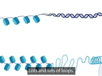
National Eye Institute researchers mapped the organization of human retinal cell chromatin, the fibers that package 3 billion nucleotide-long DNA molecules into compact structures that fit into chromosomes within each cell’s nucleus. The resulting comprehensive gene regulatory network provides insights into regulation of gene expression in general, and in retinal function, in both rare and common eye diseases. The study published in Nature Communications.
Black patients have a dramatically higher risk of advanced vision loss after a new diagnosis of primary open angle glaucoma (POAG) when compared to white patients, according to a new study from New York Eye and Ear Infirmary of Mount Sinai (NYEE).
Johns Hopkins Medicine researchers have developed a color-coded test that quickly signals whether newly developed nanoparticles — ultra small compartments designed to ferry medicines, vaccines and other therapies — deliver their cargo into target cells. The new testing tool, engineered specifically to test nanoparticles, could advance the search for next-generation biological medicines.
Researchers at Washington University School of Medicine in St. Louis have received two grants from the National Institutes of Health (NIH) totaling more than $5.5 million to develop new treatments for two types of devastating parasitic infections common in sub-Saharan Africa and Central and South America: river blindness and intestinal worm infections.
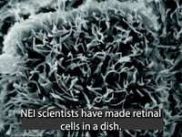
Scientists have discovered that gene therapy and the diabetes drug metformin may be potential treatments for late-onset retinal degeneration (L-ORD), a rare, blinding eye disease. Researchers from the National Eye Institute (NEI), part of the National Institutes of Health generated a “disease-in-a-dish” model to study the disease. The findings are published in Communications Biology.
As regenerative therapies for blinding diseases move closer to clinical trials, the National Eye Institute’s functional imaging consortium, a part of the NEI Audacious Goals Initiative (AGI), is pioneering noninvasive technologies to monitor the function of the retina’s light-sensing neurons and their connections to the brain.
Researchers at the University of Illinois Chicago have been awarded a five-year, $10.15 million grant to develop a broad-spectrum immunomodulatory eye drop.
A high-sugar diet creates a ‘double jeopardy’ impact for a protein crucial to cellular housekeeping, a new study suggests. The protein offsets cell damage from sugar, but too much sugar renders it ineffective. The results may offer insight for reducing age-related degenerative disease.
Shantanu Chakrabartty’s laboratory has been working to create sensors that can run on the least amount of energy. His lab has been so successful at building smaller and more efficient sensors, that they’ve run into a roadblock in the form of a fundamental law of physics.Sometimes, however, when you hit what appears to be an impenetrable roadblock, you just have to turn to quantum physics and tunnel through it.
The NIH grant will enable FAU scientists to identify the gene regulation pathways activated to program immature stem-like cells of the eye lens to attain their mature form and transparent function. The research team plans to explore the genetic and cellular mechanisms controlling developmental DNA conformational changes and will identify the transcription factors needed for eye lens formation.
Scientists from the John A. Moran Eye Center at the University of Utah have achieved another first in the field of connectomics, which studies the synaptic connections between neurons. The National Institutes of Health (NIH)-funded lab has produced the first pathoconnectome, showing how eye disease alters retinal circuitry.
Surgeons at Mass Eye and Ear have replaced the ocular surface of four patients who each experienced chemical burns to one eye by using their own stem cells taken from the other healthy eye, in a technique known as “cultivated autologous limbal epithelial cell transplantation” (CALEC). These four cases, all part of an ongoing clinical trial supported by the National Eye Institute of the NIH, represent the first procedures of their kind to occur in the United States.
Children wearing multifocal contact lenses had slower progression of their myopia, according to results from a clinical trial funded by the National Eye Institute, part of the National Institutes of Health. The findings support an option for controlling the condition, also called nearsightedness, which increases the risk of cataracts, glaucoma and retinal detachment later in life. Investigators of the Bifocal Lenses In Nearsighted Kids (BLINK) Study published the results August 11 in the Journal of the American Medical Association.
Cerebral (cortical) visual impairment (CVI) is a condition that interferes with the ability of the brain to process information from the eyes, and it has become a leading cause of visual impairment in the U.S.
Exercise can slow or prevent the development of macular degeneration and may benefit other common causes of vision loss, such as glaucoma and diabetic retinopathy, new research suggests.
Babies born prematurely who require treatment to prevent blindness from retinopathy of prematurity (ROP) could be treated with a dose of Avastin (bevacizumab) that is a fraction of the dose commonly used for ROP currently. Results from the dose-finding study were published April 23 in JAMA Ophthalmology. The study was conducted by the Pediatric Eye Disease Investigator Group (PEDIG) and supported by the National Eye Institute (NEI), part of the National Institutes of Health.
National Eye Institute (NEI) researchers profiling epigenomic changes in light-sensing mouse photoreceptors have a clearer picture of how age-related eye diseases may be linked to age-related changes in the regulation of gene expression. The findings, published online April 21 in Cell Reports, suggest that the epigenome could be targeted as a therapeutic strategy to prevent leading causes of vision loss, such as age-related macular degeneration (AMD).
Johns Hopkins Medicine scientists say they have successfully turned back the biological hands of time, coaxing adult human cells in the laboratory to revert to a primitive state, and unlocking their potential to replace and repair damage to blood vessels in the retina caused by diabetes. The findings from this experimental study, they say, advance regenerative medicine techniques aimed at reversing the course of diabetic retinopathy and other blinding eye diseases.
In a recent study using mice, lab-grown human retinal cells and patient samples, Johns Hopkins Medicine scientists say they found evidence of a new pathway that may contribute to degeneration of the light sensitive tissue at the back of the eye. The findings, they conclude, bring scientists a step closer to developing new drugs for a central vision-destroying complication of diabetes that affects an estimated 750,000 Americans.
Scientists have long sorted cells into different varieties based on their appearance under a microscope or, for differences that are more visually subtle, based on the behavior of a handful of genes. But in a bid to reveal even more distinctive differences and similarities, researchers from the Johns Hopkins Kimmel Cancer Center, the Johns Hopkins Institute for Genetic Medicine and the Johns Hopkins Department of Neuroscience developed two new artificial intelligence methods that decipher complex gene activity controlling cell fate decisions in retina development and relate this gene activity to what occurs in other tissues and across different species.
Johns Hopkins Medicine scientists say that new experiments with mouse eye tissues strongly suggest that a longstanding “textbook concept” about the way a mammal’s retina processes light needs a rewrite.
In studies with lab-grown human cells and in mice, Johns Hopkins Medicine researchers have found that an experimental drug may be twice as good at fighting vision loss as previously thought.
The process, discovered in the axons of neurons, is implicated in Alzheimer’s, amyotrophic lateral sclerosis, traumatic brain injury and other diseases or injuries to the nervous system.
Graphene electrodes could enable higher quality brain imaging thanks to new research by a team of engineers and neuroscientists at UC San Diego. The researchers developed a technique, using platinum nanoparticles, to lower the impedance of graphene electrodes by 100 times while keeping them transparent. In tests on transgenic mice, the electrodes were able to record and image neuronal activity (calcium ion spikes) at of large groups of neurons and individual brain cells.
Tim Goetz drives about 200,000 miles each year. Remarkably, Goetz is legally blind. Research funded by the National Eye Institute (NEI) is helping Goetz and others like him get or stay behind the wheel while keeping roads safe for everyone.
Common, unavoidable eye movements may be a cause of glaucoma in people with normal intraocular pressure (normal-tension glaucoma), according to new research supported by the National Eye Institute. The findings suggest that over time eye movement strains the optic nerve, the bundle of nerve fibers between the eye and brain. The research may also explain why tension-lowering eye drops can improve normal-tension glaucoma. Glaucoma is the second leading cause of blindness worldwide and January is Glaucoma Awareness Month.
Florida researchers have identified a signaling pathway that is essential for angiogenesis, the growth of new blood vessels from pre-existing vessels. The findings, published in Nature Communications, may improve current strategies to improve blood flow in ischemic tissue, such as that found in atherosclerosis and peripheral vascular disease associated with diabetes.
The eyes are for seeing, but they have other important biological functions, including automatic visual reflexes that go on without awareness. The reflexive system of the human eye also produces a conscious, visual experience, according to a new study from researchers in the Perelman School of Medicine and School of Arts and Sciences at the University of Pennsylvania.
Researchers at University of Utah Health have identified a protein (ARF6) that when inhibited reduces diabetic retinopathy, a condition that results when blood vessels at the back of the eye leak fluid into the eye, impairing vision.
A protein shaped like a "Y" makes scientists do a double-take and may change the way they think about a protein sometimes implicated in glaucoma. The Y is a centerpiece in myocilin, binding four other components nicknamed propellers together like balloons on strings.
Several studies indicate that the prevalence of myopia is increasing in the U.S. and worldwide, and researchers project that the trend will continue in the coming decades. Otherwise known as nearsightedness, myopia occurs when the eye grows too long from front to back. Instead of focusing images on the retina—the light-sensitive tissue in the back of the eye—images are focused at a point in front of the retina. As a result, people with myopia have good near vision but poor distance vision.
Cytokines might be the key to repairing diabetic nerve damage. Diabetes devastates nerve cells, which can lead to poor circulation, muscle weakness, blindness, and other side effects. The study showed diabetic mice can’t repair nerve cells after damage due to low levels of specific cytokines.
Cells within an injured mouse eye can be coaxed into regenerating neurons and those new neurons appear to integrate themselves into the eye’s circuitry, new research shows. The findings potentially open the door to new treatments for eye trauma and retinal disease. The study appears in the July 26 issue of Nature, and was funded in part by the National Eye Institute (NEI), part of the National Institutes of Health.
A Wayne State University researcher recently received a $1.9 million grant from the National Eye Institute of the National Institutes of Health for the project, “Role of AMP-Activated Protein Kinase in Bacterial Endophthalmitis.”
Endophthalmitis is a severe inflammation of the interior of the eye caused by contaminating microorganisms that enter the eye following trauma or surgery, or that spread through the bloodstream from a distant infection site. Despite appropriate therapeutic intervention, bacterial endophthalmitis often results in vision loss and sometimes requires surgical removal of the eye.
Bugs in your eyes may be a good thing. Resident microbes living on the eye are essential for immune responses that protect the eye from infection, new research shows. The study, which appears in the journal Immunity on July 11, demonstrates the existence of a resident ocular microbiome that trains the developing immune system to fend off pathogens. The research was conducted at the National Eye Institute (NEI), part of the National Institutes of Health.
Wayne State University recently received a five-year, $1.925 million grant from the National Eye Institute of the National Institutes of Health to test the role of microRNAs (miRNAs) — a newly recognized level of gene expression regulation — in bacterial keratitis – an infection of the cornea caused by bacteria — as well as to identify new therapeutic targets and alternative treatment strategies.
Monthly eye injections of Avastin (bevacizumab) are as effective as the more expensive drug Eylea (aflibercept) for the treatment of central retinal vein occlusion (CRVO), according to a clinical trial funded by the National Eye Institute (NEI), part of the National Institutes of Health. After six monthly injections, treatment with either drug improved visual acuity on average from 20/100 to 20/40.
Systemic therapy consisting of corticosteroids and immunosuppressants preserved vision of uveitis patients better – and had fewer adverse outcomes – than a long-lasting corticosteroid intraocular implant, according to a clinical trial funded by the National Eye Institute (NEI). After seven years, visual acuity on average remained stable among participants on systemic therapy but declined by an average of six letters (about one line on an eye chart) among participants who had the implant. NEI is part of the National Institutes of Health.
In experiments with a protein called Ephexin5 that appears to be elevated in the brain cells of Alzheimer's disease patients and mouse models of the disease, Johns Hopkins researchers say removing it prevents animals from developing Alzheimer's characteristic memory losses. In a report on the studies, published online March 27 in The Journal of Clinical Investigation, the researchers say the findings could eventually advance development of drugs that target Ephexin5 to prevent or treat symptoms of the disorder.