
Newswise — Materials — Molding molecular matter
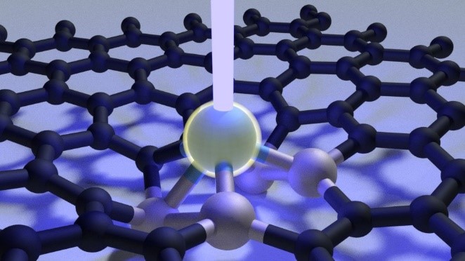
Scientists at Oak Ridge National Laboratory used a focused beam of electrons to stitch platinum-silicon molecules into graphene, marking the first deliberate insertion of artificial molecules into a graphene host matrix.
While scientists have already used the beam of a high-resolution electron microscope to intentionally rearrange graphene’s carbon-based molecular structure, this new development greatly enhances scientists’ ability to control matter at the atomic scale.
“This technique allows us to insert foreign molecules into the graphene lattice to change its physical properties,” said ORNL’s Ondrej Dyck.
He explained that this process is generally applicable and could be especially useful for prototyping quantum-based devices — including solid state qubits for quantum computers — from graphene and other ultra-thin materials.
The research was published in Carbon. – Gage Taylor
Image: https://www.ornl.gov/sites/default/files/2020-04/Ebeam_IMAGE_Final.jpg
Caption: Researchers insert a platinum-silicon molecule into a graphene lattice with a focused electron beam. Credit: Ondrej Dyck/Oak Ridge National Laboratory, U.S. Dept. of Energy
Nuclear — Seeing inside particles
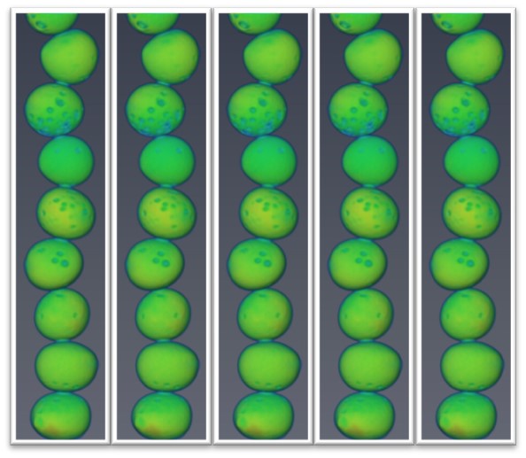
Oak Ridge National Laboratory researchers working on neutron imaging capabilities for nuclear materials have developed a process for seeing the inside of uranium particles – without cutting them open.
Nuclear materials experts and neutron scientists collaborated on the process, which creates image-based 3D reconstructions of the particles. Researchers can even view “slices” of different layers of particles to learn the effects of various conditions on elemental distribution, density and other properties.
Other methods to characterize these particles require they be cut in half, put in epoxy and mounted for viewing with an electron microscope. The new technique allows for characterization of the entire particle, not just a single cross section. ORNL’s Kristian Myhre said the process is broadly applicable and already is being used to study other materials.
“Unlike electron microscopy, the neutron imaging allows you to take a unique picture without destroying your sample,” Myhre said. “It’s such a versatile technique.”
Image: https://www.ornl.gov/sites/default/files/2020-04/Kernels-nuclear%20materials-2.jpg
Caption: Neutron imaging can non-destructively view important characteristics to develop advanced nuclear materials, such as the overall shape and defects shown in this example 3D image of uranium TRISO kernels. Credit: Kristian Myhre/Oak Ridge National Laboratory, U.S. Dept. of Energy