Loss of the protein pigment epithelium-derived factor (PEDF), which protects retinal support cells, may drive age-related changes in the retina, according to a new study in mice from the National Eye Institute (NEI).
Loss of the protein pigment epithelium-derived factor (PEDF), which protects retinal support cells, may drive age-related changes in the retina, according to a new study in mice from the National Eye Institute (NEI).
Clinical trial results from the DRCR Retina Network suggest that a specific step strategy, in which patients with diabetic macular edema start with a less expensive medicine and switch to a more expensive medicine if vision does not improve sufficiently, gives results similar to starting off with the higher-priced drug.
The eye’s light-sensing retina taps different circuits depending on whether it is generating image-forming vision or carrying out a non-vision function such as regulating pupil size or sleep/wake cycles, according to a new mouse study from the National Eye Institute (NEI) and the National Institute of Mental Health (NIMH).
The eye’s light-sensing retina taps different circuits depending on whether it is generating image-forming vision or carrying out a non-vision function such as regulating pupil size or sleep/wake cycles, according to a new mouse study from the National Eye Institute (NEI) and the National Institute of Mental Health (NIMH).
In a study of eye fluid from 38 patients, Johns Hopkins Medicine researchers say they have found that levels of a specific protein appears to help accurately predict whether people with the wet form of age-related macular degeneration may need lifelong, frequent eye injections to preserve vision or if they can be safely weaned off the treatments.
Byron Lam and collaborators at the University of Miami reported results from an 8-patient phase 1 gene therapy clinical trial for the degenerative retinal disease Leber hereditary optic neuropathy. They found no significant safety concerns; however, treatment failed to improve or slow vision loss, with even the highest dose.
Researchers have identified distinct differences among the cells comprising a tissue in the retina that is vital to human visual perception. The scientists from the National Eye Institute (NEI) discovered five subpopulations of retinal pigment epithelium (RPE)—a layer of tissue that nourishes and supports the retina’s light-sensing photoreceptors. Using artificial intelligence, the researchers analyzed images of RPE at single-cell resolution to create a reference map that locates each subpopulation within the eye.
Researchers have identified distinct differences among the cells comprising a tissue in the retina that is vital to human visual perception. The scientists from the National Eye Institute (NEI) discovered five subpopulations of retinal pigment epithelium (RPE)—a layer of tissue that nourishes and supports the retina’s light-sensing photoreceptors. Using artificial intelligence, the researchers analyzed images of RPE at single-cell resolution to create a reference map that locates each subpopulation within the eye.
New research shows that a treatment for retinal vein occlusion yields long-lasting vision gains, with visual acuity remaining significantly above baseline at five years. However, many patients require ongoing treatment.
The road from discovering a potential drug to getting the therapy into the hands of patients is a long and uncertain one. An NIH program offers researchers a smoother path from basic science to clinical testing and regulatory approval.
The road from discovering a potential drug to getting the therapy into the hands of patients is a long and uncertain one. An NIH program offers researchers a smoother path from basic science to clinical testing and regulatory approval.
New research by National Eye Institute (NEI) investigators shows that while microsaccades seem to boost or diminish the strength of brain signals underlying attention, the eye movements are not drivers of those brain signals.
New research by National Eye Institute (NEI) investigators shows that while microsaccades seem to boost or diminish the strength of brain signals underlying attention, the eye movements are not drivers of those brain signals.
National Eye Institute researchers developed and validated an artificial-intelligence-based method to evaluate patients with Stargardt, an eye disease that can lead to childhood vision loss. The method quantifies disease-related loss of light-sensing retina cells, yielding information for monitoring patients, understanding genetic causes of the disease, and developing therapies to treat it.
Johns Hopkins Medicine researchers have developed a color-coded test that quickly signals whether newly developed nanoparticles — ultra small compartments designed to ferry medicines, vaccines and other therapies — deliver their cargo into target cells. The new testing tool, engineered specifically to test nanoparticles, could advance the search for next-generation biological medicines.
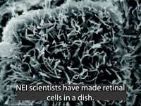
Using a stem-cell-derived model, researchers have identified two drug candidates that may slow dry age-related macular degeneration (AMD), a leading cause of blindness for which no treatment exists. The scientists, from the National Eye Institute (NEI), part of the National Institutes of Health, published their findings today in Nature Communications.
Scientists have discovered that gene therapy and the diabetes drug metformin may be potential treatments for late-onset retinal degeneration (L-ORD), a rare, blinding eye disease. Researchers from the National Eye Institute (NEI), part of the National Institutes of Health generated a “disease-in-a-dish” model to study the disease. The findings are published in Communications Biology.
Using data generated from patients and mice with genetic mutation for the disorder Usher syndrome, researchers from the University of Maryland School of Medicine and the National Institutes of Health documented the natural history of vision impairment in patients and identified the cell mechanism behind progressive vision loss.
A new study by University of Illinois Chicago researchers shows a mechanism that stops the herpes simplex virus 1 from causing serious brain damage and death. Researchers discovered a function of a protein complex, mammalian target of rapamycin complex 2, in an antiviral defense mechanism. This protein complex limits HSV-1 virus infection through rapid activation of antiviral immunity and protects the host by preventing encephalitis — brain inflammation — and possible death due to HSV-1 infection.
A new study by researchers at University of Illinois Chicago suggests that when the protein optineurin, or OPTN, is present in cells it restricts the spread of HSV-1, the herpes simplex virus type 1.In a “first of its kind” study, researchers also found a potential direct connection between neurodegenerative diseases, such as Alzheimer’s disease, amyotrophic lateral sclerosis (ALS), glaucoma, and the herpesvirus.
Equipped with a color 3D camera, an inertial measurement sensor, and its own on-board computer, a newly improved robotic cane could offer blind and visually impaired users a new way to navigate indoors.
Researchers at the National Institutes of Health have discovered that decisions based on visual information, which involve a complex stream of data flowing forward and backwards along the brain’s visual pathways, is broadcast widely to neurons in the visual system, including to those that are not being used to make the decision.
Researchers at the National Institutes of Health have discovered that decisions based on visual information, which involve a complex stream of data flowing forward and backwards along the brain’s visual pathways, is broadcast widely to neurons in the visual system, including to those that are not being used to make the decision.
A form of gene therapy protects optic nerve cells and preserves vision in mouse models of glaucoma, according to research supported by NIH’s National Eye Institute. The findings suggest a way forward for developing neuroprotective therapies for glaucoma, a leading cause of visual impairment and blindness.
A form of gene therapy protects optic nerve cells and preserves vision in mouse models of glaucoma, according to research supported by NIH’s National Eye Institute. The findings suggest a way forward for developing neuroprotective therapies for glaucoma, a leading cause of visual impairment and blindness.
The vision community and its coalition partners announce awareness and educational activities in July 2021 around the annual recognition of Dry Eye Awareness Month.
Scientists studied the brain activity of school-aged children during development and found that regions that activated upon seeing limbs (hands, legs, etc.) subsequently activated upon seeing faces or words when the children grew older. The research, by scientists at Stanford University, Palo Alto, California, reveals new insights about vision development in the brain and could help inform prevention and treatment strategies for learning disorders. The study was funded by the National Eye Institute and is published in Nature Human Behaviour.
Scientists studied the brain activity of school-aged children during development and found that regions that activated upon seeing limbs (hands, legs, etc.) subsequently activated upon seeing faces or words when the children grew older. The research, by scientists at Stanford University, Palo Alto, California, reveals new insights about vision development in the brain and could help inform prevention and treatment strategies for learning disorders. The study was funded by the National Eye Institute and is published in Nature Human Behaviour.
Researchers at the National Eye Institute (NEI) have determined how certain short protein fragments, called peptides, can protect neuronal cells found in the light-sensing retina layer at the back of the eye. The peptides might someday be used to treat degenerative retinal diseases, such as age-related macular degeneration (AMD).
Researchers at the National Eye Institute (NEI) have determined how certain short protein fragments, called peptides, can protect neuronal cells found in the light-sensing retina layer at the back of the eye. The peptides might someday be used to treat degenerative retinal diseases, such as age-related macular degeneration (AMD).
What exactly triggers a sneeze? A team led by researchers at Washington University School of Medicine in St. Louis has identified, in mice, specific cells and proteins that control the sneeze reflex. Better understanding of what causes us to sneeze — specifically how neurons behave in response to allergens and viruses — may point to treatments capable of slowing the spread of infectious respiratory diseases.
In a new study, supported by a five-year, $6.4 million grant from the National Eye Institute of the National Institutes of Health, researchers from Case Western Reserve University, University Hospitals and the Jaeb Center for Health Research, aim to finally determine which diabetic individuals can successfully donate their corneas for keratoplasty (and which should not).
As regenerative therapies for blinding diseases move closer to clinical trials, the National Eye Institute’s functional imaging consortium, a part of the NEI Audacious Goals Initiative (AGI), is pioneering noninvasive technologies to monitor the function of the retina’s light-sensing neurons and their connections to the brain.
NIH supported early testing of the artificial retina. Now, scientists are testing whether manufacturing it on the International Space Station results in a viable treatment for people with blinding eye diseases.
Early treatment with anti-VEGF injections slowed diabetic retinopathy in a clinical study from the DRCR Retina Network (DRCR.net). However, two years into the four-year study its effect on vision was similar to standard treatment, which usually begins at the onset of late disease.
Early treatment with anti-VEGF injections slowed diabetic retinopathy in a clinical study from the DRCR Retina Network (DRCR.net). However, two years into the four-year study its effect on vision was similar to standard treatment, which usually begins at the onset of late disease.
The National Eye Institute (NEI) Data Commons now enables researchers to access data from patients with macular degeneration who participated in the Age-related Eye Disease Study 2 (AREDS2). The database complements newly available stem cell lines created by the New York Stem Cell Foundation Research Institute (NYSCF) from blood cells of AREDS2 study participants.
In the largest genome-wide association study of glaucoma to date, an international team of researchers compared the genes of 34,179 people with the disease to 349,321 control subjects. They identified 127 genes linked to glaucoma, including 44 new gene loci and confirmed 83 previously reported loci.
Working with fish, birds and mice, Johns Hopkins Medicine researchers report new evidence that some animals’ natural capacity to regrow neurons is not missing, but is instead inactivated in mammals. Specifically, the researchers found that some genetic pathways that allow many fish and other cold-blooded animals to repair specialized eye neurons after injury remain present in mammals as well, but are turned off, blocking regeneration and healing.
Scientists at the National Eye Institute (NEI) have developed a promising gene therapy strategy for a rare disease that causes severe vision loss in childhood. A form of Leber congenital amaurosis, the disease is caused by autosomal-dominant mutations in the CRX gene, which are challenging to treat with gene therapy.
Scientists at the National Eye Institute (NEI) have developed a promising gene therapy strategy for a rare disease that causes severe vision loss in childhood. A form of Leber congenital amaurosis, the disease is caused by autosomal-dominant mutations in the CRX gene, which are challenging to treat with gene therapy.
The Association for Research in Vision and Ophthalmology (ARVO) today announced the recipient of the Mallinckrodt Uveitis Research Fellowship, generously funded by Mallinckrodt Pharmaceuticals. The award, funded through the ARVO Foundation, supports an early career investigator with a one-year grant of $45,000 to study an aspect of uveitis or other inflammatory conditions of the eye. The 2021 awardee is Shilpa Kodati, MD of the National Eye Institute (NEI) at the National Institutes of Health (NIH).
In experiments in mouse tissues and human cells, Johns Hopkins Medicine researchers say they have found that removing a membrane that lines the back of the eye may improve the success rate for regrowing nerve cells damaged by blinding diseases. The findings are specifically aimed at discovering new ways to reverse vision loss caused by glaucoma and other diseases that affect the optic nerve, the information highway from the eye to the brain.
Researchers at the University of Illinois Chicago have published a study showing a promising approach to using drug repurposing to treat genetic diseases. A team from the UIC Department of Ophthalmology and Visual Sciences published the article, “Gene dosage manipulation alleviates manifestations of hereditary PAX6 haploinsufficiency in mice” in the journal Science Translational Medicine.
Researchers at the National Eye Institute (NEI) report that a brain region in the superior temporal sulcus (fSTS) is crucial for processing and making decisions about visual information.
Researchers at the National Eye Institute (NEI) report that a brain region in the superior temporal sulcus (fSTS) is crucial for processing and making decisions about visual information.
Surgical and injectable drug approaches are equally effective for treatment of bleeding inside the eye from proliferative diabetic retinopathy (PDR), according to a National Eye Institute (NEI)-supported clinical study from the DRCR Retina Network (DRCR.net).